
Dental Care for Pets
As a veterinary dental practice, we focus particularly on the dental health of our patients.
Herbivorous species (rabbits, guinea pigs, chinchillas, degus) are increasingly being brought in with dental problems. What sets them apart from other rodents is that even their molars lack true tooth roots and continue to grow throughout their lives. Unfortunately, over the course of their lives, congenital misalignments or acquired misalignments —caused by inflammation, for example—can develop, altering the way the teeth grow. As a result, the molars of the upper and lower jaws no longer fit together perfectly, leading to a missing or imperfect bite. When the chewing surfaces of the upper and lower jaws no longer align perfectly, the teeth can no longer grind food as effectively, causing them to continue growing. A vicious cycle ensues .
The sooner you intervene, the better you can prevent more serious problems. However, existing misalignment of the teeth cannot be corrected. Tooth extraction is possible and advisable in some cases; however, these herbivorous species are heavily dependent on their dentition and functional anatomy. For this reason, it is unfortunately not possible to extract all their teeth, as is done, for example, with carnivores such as dogs and cats. Rabbits and guinea pigs would then no longer be able to eat or engage in activities appropriate to their species. A permanent diet of pureed food is not recommended for animal welfare reasons.
Tooth correction—that is, the grinding down of chewing surfaces in the molar region—must always be performed under anesthesia. Otherwise, defensive movements against the instruments or chewing and biting motions against the instruments held in place within the oral cavity could result in new tooth damage and oral cavity injuries. Simply clamping down on the molars carries the risk of tooth fractures. For this reason, modern veterinary medicine uses electric grinding instruments. These ensure a finer, more even result and are significantly safer.
Before orthodontic treatment, various X-rays should always be taken. We take these images using our dental X-ray machine. They reveal the portion of the teeth—which accounts for as much as 80%—that lies within the jawbone and is therefore not visible during an examination of the oral cavity. Here, we can identify abscesses, elongated teeth, widened teeth (macrodontia in guinea pigs), and tooth rotations (rotation around the tooth axis). In addition, measurement methods applied to the X-ray image—by marking on the image—allow us to determine the necessary correction, that is, the portion of the teeth that must be ground down (according to Böhmer and Crowley).
In some cases, misalignment limited to the incisors can be corrected without anesthesia. Misalignment of the incisors can also occur in species that primarily eat grains; in such cases, anesthesia may be advisable due to the animal’s size.
Small carnivores, such as ferrets, receive the same dental care at our clinic as dogs and cats. Dental cleanings are performed under anesthesia. During the procedure, patients are intubated, kept warm, and monitored. After tartar removal using a scaler, the teeth are probed (a blunt probe is used to check whether the tooth necks are exposed and the periodontal pockets are enlarged), X-rayed, and extracted if necessary.
Our dental X-rays can reveal changes in the teeth, the alveolar socket, and the bone (e.g., abscesses). Only after taking dental X-rays can we reliably determine which teeth need to be extracted. Finally, the teeth are polished to ensure the surface is nice and smooth, preventing new tartar from immediately forming on the rough surface.
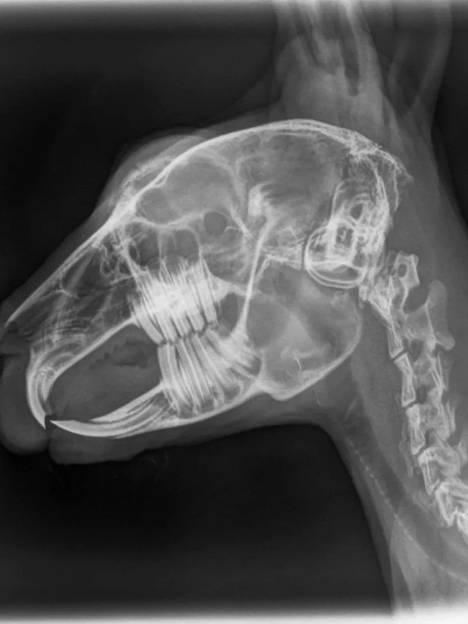
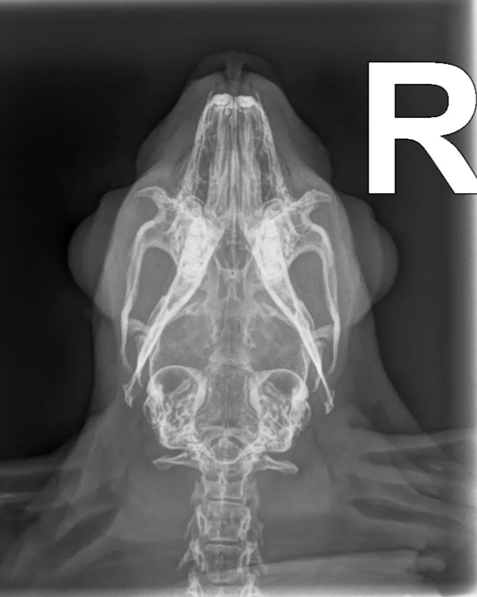
Photo: X-ray image of a rabbit's head