
Tooth-Preserving Surgery
Options for preserving teeth in dogs and cats based on the principles of modern oral surgery at the Sonnenallee Veterinary Practice in Berlin-Neukölln.
When Is It Worth It to Save a Tooth?
Not every damaged or inflamed tooth needs to be extracted. It is possible to save a tooth instead of extracting it. If the tooth structure is largely intact and the surrounding jawbone provides sufficient support, we have specialized procedures at our disposal:
Dental Fillings: Whether due to accidents, wear and tear, or as part of cavity prevention, modern filling materials protect exposed dentin from irritants and bacteria.
Endodontics & Root Canal Treatment: In cases of deep tooth fractures or dead tissue, we thoroughly clean and disinfect the root canal before sealing it tightly. This ensures that the tooth remains firmly anchored in the jaw.
Vital pulp therapy: In the case of a fresh fracture (usually less than 48 hours old) in a young animal, the living nerve tissue (pulp) can often be saved by placing a crown over the tooth, allowing it to continue growing normally.
Periodontal Surgery: In cases of advanced periodontitis-related gum recession, we surgically clean deep gum pockets, remove inflamed tissue, and use special preparations to promote the regeneration of the periodontal tissues.
All procedures at our veterinary practice in Berlin-Neukölln are performed under gentle, closely monitored general anesthesia and using digital dental X-rays to precisely assess the extent of the problem beneath the gums. Your pet will recover quickly after the procedure: With appropriate pain management and a temporary soft-food diet, the healing process is usually uncomplicated.
Have you noticed a broken tooth or inflammation in your pet? Bring your pet in for a dental exam at our practice in Berlin-Neukölln so we can discuss the best option for saving the tooth.
Periodontal Surgery
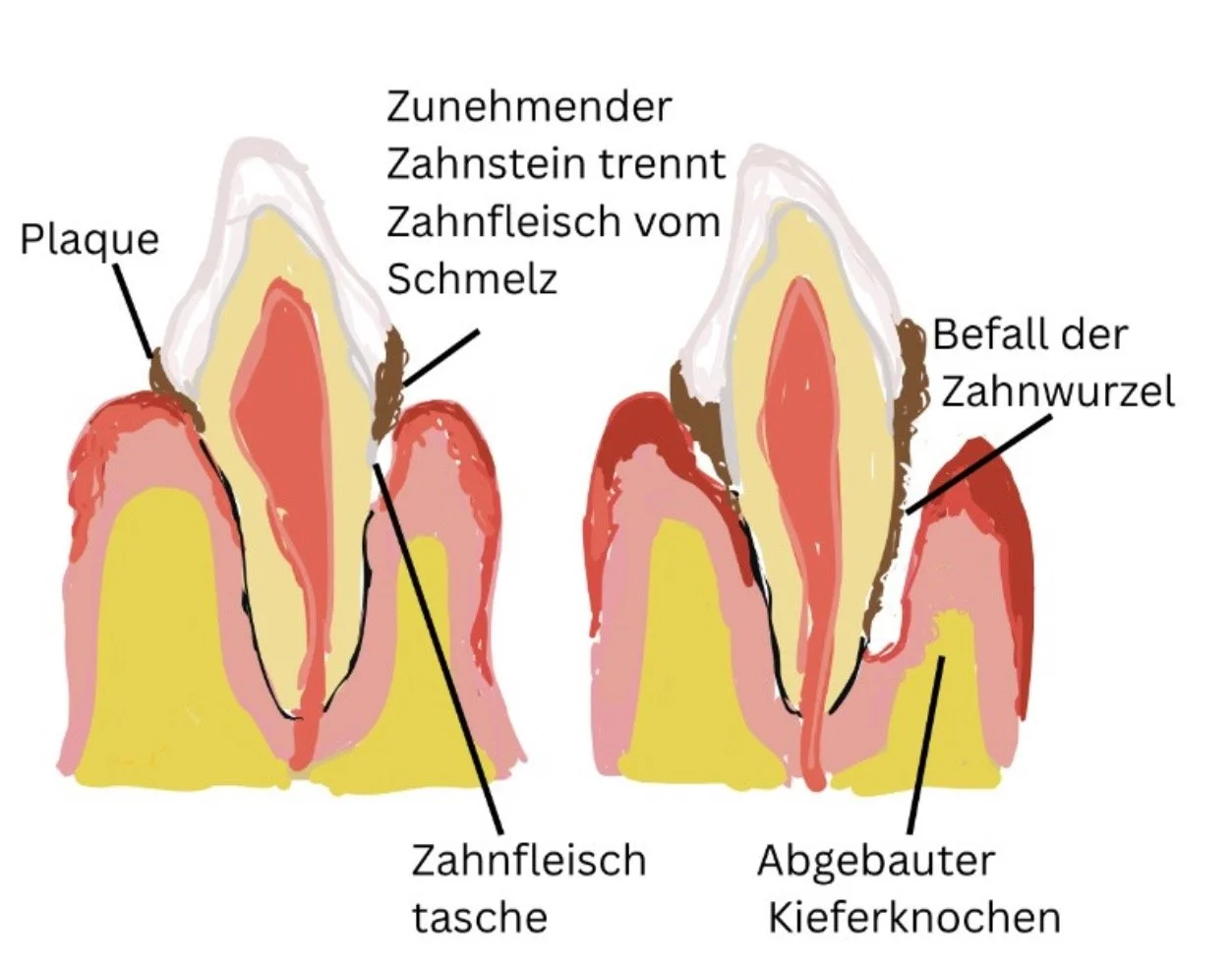
Periodontal surgery is used, among other things, when a simple professional dental cleaning is no longer sufficient to save the periodontium (the structures supporting the teeth). The goal is always to eliminate deep-seated bacterial colonies, stop the breakdown of the jawbone, and, in the best-case scenario, rebuild the tissue.
The Most Important Procedures in Periodontal Surgery
Open Wound Debridement (Flap Surgery / Open Flap Debridement): In cases of deep gum pockets (more than 3–4 mm in dogs), external access is no longer sufficient. The gum tissue is carefully surgically lifted away from the bone (flap surgery). Only then do deep-seated calculus, tartar, and infected tissue around the tooth roots become visible, allowing them to be thoroughly removed under direct visualization and the tooth root to be smoothed. The gum tissue is then sutured back into place.
Guided Tissue Regeneration (GTR): If periodontitis has already led to localized bone loss, this procedure protects the periodontal tissues. After cleaning the pocket, special resorbable membranes are placed in combination with bone substitute material. They prevent the rapidly growing gum tissue from invading the defect and allow the slower-growing bone and periodontal ligament tissue time to regenerate.
Gingivectomy & Gingivoplasty: Some animals—particularly dogs of certain breeds or cats with chronic inflammation—are prone to gum overgrowth (gingival hyperplasia). Bacteria accumulate extremely easily in these deep gum pockets. Surgical removal (gingivectomy) and reshaping (gingivoplasty) of the excess tissue eliminate these pockets and make cleaning easier again.
Mucogingival surgery: In cases of exposed roots or tissue defects (e.g., oronasal fistulas—a connection between the oral and nasal cavities resulting from severe periodontal damage to the canines), healthy mucosal tissue is repositioned and sutured to permanently close the defect.
Endodontics and Vital Pulp Therapy
The main difference lies in the condition of the dental nerve (the pulp): While root canal treatment involves the complete removal of the inflamed or dead tissue, vital pulp therapy aims to preserve the dental nerve.
Root Canal Treatment (Endodontics)
Condition of the nerve: dead , severely inflamed, or infected
Purpose of the procedure: To preserve a tooth in the jaw even though it is "dead"
Procedure: The root canal is completely cleaned out, disinfected, and sealed.
Common causes: Older tooth fractures; teeth that have died as a result of trauma or infection (often appear darker).
Age of the animal: Mostly adult animals whose teeth have finished growing.
Vital Pulp Therapy
Condition of the nerve: alive , healthy, or only slightly irritated
Goal of the procedure: To fully preserve the vitality (life) of the tooth
Procedure: The tissue is disinfected, covered with a protective agent, and sealed.
Typical cause: Recent tooth fractures (ideally within 24–48 hours), planned tooth reduction, e.g., due to congenital malocclusion.
Age of the animal: This is especially important for young animals (under 1.5–2 years old).
Why is this difference so important for young animals?
In young animals, the tooth wall is still very thin and the root apex is open. If the nerve dies, the growth of the tooth wall stops, and the tooth remains thin and unstable. Vital pulp therapy is the treatment of choice at this age, since a living nerve ensures that the tooth wall continues to thicken and the root matures fully.
However, if the tooth has been broken for some time or bacteria have already entered it, root canal treatment is the safest option for preventing infection in the jawbone while still preserving the tooth and keeping it pain-free.
Root Canal Treatment - Endodontics
Root canal treatment (endodontics) in dogs and cats is performed to save a strategically important tooth—usually the largecanines or thefourth premolar—when its nerve is infected, dead, or exposed following an accident.Instead of extracting the tooth, the inflamed tissue inside is completely removed, the cavity is disinfected, and the tooth is sealed tightly.
The Process of a Root Canal Treatment
The procedure is performed under monitored general anesthesia and local anesthesia. Digital dental X-rays are used to assess the root structure. This involves checking whether the root tip is already damaged or whether, for example, there are any longitudinal fractures that would make preservation impossible.
The tooth is then drilled from the top or the side to create direct access to the pulp canal (the nerve canal). Using specialized, flexible endodontic files, the dead or inflamed nerve and vascular tissue is precisely removed all the way to the root tip.
The root canal is shaped in several steps using special files and is thoroughly rinsed with antibacterial solutions in between to eliminate all bacteria and tissue debris. After thorough cleaning and disinfection, the root canal is dried.
To prevent bacteria from re-entering through the jaw or the trepanation site, the cavity is sealed with a precise fit using a combination of a special sealing paste (sealer) and biocompatible rods (guttapercha).
The drilled opening is then sealed with a multilayer filling (e.g., adhesive resin/composite) to make it load-bearing and liquid-tight.
X-ray examinations:
Throughout the entire treatment process, several X-rays are necessary to confirm the correct depth of the files in the root canal and that the canal has been completely filled all the way to the root tip.
Important Prerequisites and Follow-Up Care
X-ray follow-up after 4–6 months and after one year: The success of the treatment must be verified by X-ray after a few months to ensure that no unnoticed inflammation has developed at the root tip.
Load-bearing capacity: The treated tooth is fully functional after the filling is placed. However, to protect the filling, you should avoid chewing very hard foods.
Vital Pulp Therapy
Vital pulp therapy refers to a tooth-preserving procedure in which the dental pulp is preserved and the tooth is restored with a crown.
Unlike root canal treatment (in which the nerve is killed and removed), the goal of this treatment is to save the living nerve tissue (pulp) inside the tooth and keep it alive.
When is vital pulp therapy an option?
For this procedure to be successful, two conditions must be met:
The tooth has recently been injured: Ideally, the fracture occurred no more than 24 to 48 hours ago.
The nerve is healthy and functioning normally: There is no deep bacterial infection or tissue necrosis yet.
This is especially important for young animals: In dogs and cats under 1.5 to 2 years of age, tooth development is not yet complete. The tooth wall is thin and the root tip is open. The tooth can continue to grow and become stable only if the nerve is alive.
The Process
A dental X-ray is taken under general anesthesia to ensure that the root is intact, that there is no longitudinal fracture of the tooth, and that the bone at the root tip has not yet been affected.
The slightly contaminated nerve tissue exposed at the site of the rupture is removed with millimeter precision at the uppermost section while being continuously cooled. The wound on the nerve is flushed clean, and the bleeding is stopped. Maintaining a sterile environment is key to success here.
A special, biocompatible material (e.g., mineral trioxide aggregate / MTA) is applied directly to the healthy, non-bleeding nerve tissue. This material soothes the nerve and stimulates it to form new protective dentin.
A base filling is applied over the protective layer, and the tooth is then sealed securely and germ-proof with a multi-layered plastic filling (composite).
Follow-up Care and Monitoring of Outcomes
Since the nerve remains in the tooth, it is essential to monitor the healing process. After 3 to 6 months, a follow-up appointment is scheduled, during which an X-ray is taken under brief anesthesia. This allows the dentist to check whether the tooth has remained viable, whether the root wall is continuing to grow as expected, and whether the surrounding bone is free of inflammation.